Breast Cancer

EPIDEMIOLOGY AND RISK FACTORS
Breast cancer is the most common malignant tumour in women, with its incidence in recent years showing an increasing trend, probably due to multiple factors including the implementation of population-based screening programs and changes in nutritional and reproductive habits. However, at the same time, breast cancer mortality has been decreasing thanks to progressive advances in oncological treatments, better knowledge of tumour biology and, above all, the establishment of these screening programs, which allow an earlier diagnosis of the disease and, therefore, the possibility to address it in its earlier stages.

OBLIQUE PROJECTIONS SHOWING A LESION IN THE LOWER QUADRANTS OF THE RIGHT BREAST (A) AND A HEALTHY LEFT BREAST (B), WITHOUT ALTERATIONS.

Although we do not have a national tumour registration system in Spain in order to know the exact figures of this disease, it is estimated that around 33,000 cases are diagnosed each year, which represents 30% of the total tumours diagnosed in women, thereby representing a significant public health problem for women.
Of these breast tumours, around 90-95% are sporadic cases, and only 5-10% of cases have an associated hereditary component. Although the cause of sporadic breast tumours is unknown, multiple risk factors that favour their development have been identified, including: age and sex, reproductive factors, endogenous and exogenous sex hormones, genetic susceptibility and lifestyle (diet, physical exercise, obesity, environmental pollution and ionizing radiation).
Various genes have been associated with a family predisposition to developing breast cancer. The two best-known genes are BRCA1 and BRCA2. The study of both genes is carried out on a patient’s blood sample and is indicated in the following situations:
  • Breast cancer diagnosed at age 45 or younger.
  • Breast cancer diagnosed between 46 and 50 years of age if accompanied by another instance of breast cancer at any age in the same patient, if the family history is not informative, or if there is at least one other instance of breast, ovary, prostate, or pancreatic cancer, diagnosed at any age, in the patient’s family history.
  • Breast cancer of the triple-negative subtype below the age of 60.
  • Breast cancer diagnosed in a male.

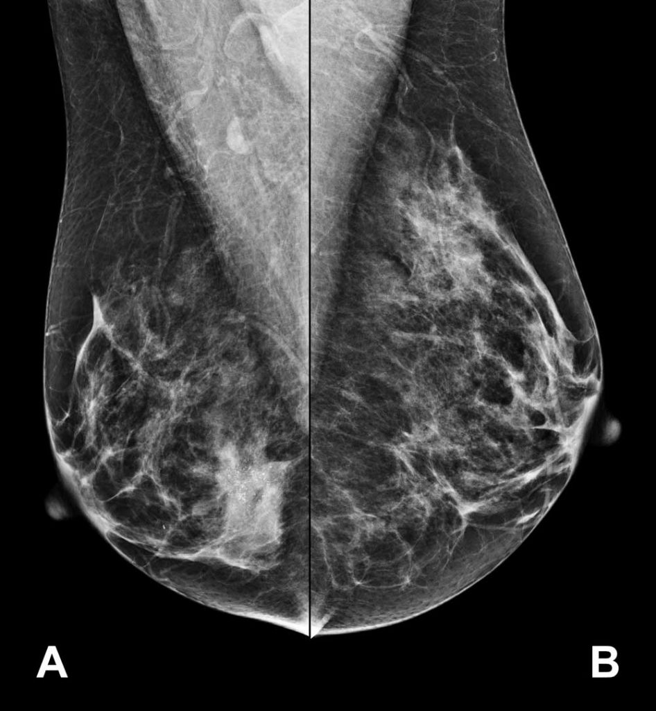
Breast cancer screening

As previously mentioned, the establishment of population-based screening programs has helped to reduce mortality from breast cancer, due to the fact that they enable diagnosis of the disease in its earlier stages. The current recommendations from the U.S. Preventive Services Task Force on breast cancer screening are:

1

To perform a mammogram every two years on women between the ages of 50 and 74.

2

The benefits of applying the screening program to women over the age of 75 are not well established.

3

Mammograms for breast cancer screening should not be given to women between the ages of 40 and 49. Their implementation in this age group should be assessed on a case-by-case basis.

4

There is insufficient current evidence to recommend scans additional to mammograms in women under the age of 40.
The recommendations for women between the ages of 40 and 49, as well as the frequency with which mammograms should be performed, have created significant controversy. Therefore there may be modifications to these general recommendations in different geographical areas, the most common alternative being the performance of an annual mammogram from the age of 40.
Several studies seem to support alternating mammograms and breast MRIs every six months in women who have an identified BRCA1 or BRCA2 mutation, as this reduces the number of breast cancer cases diagnosed in advanced stages. However, the use of breast MRI outside this group of patients is not recommended.

DETECTION

The clinical manifestation of breast cancer varies according to its extent and location in the breast, as well as the presence or absence of distant metastases.
It was initially thought that, after a variable period of growth within the mammary gland, the primary tumour invaded the locoregional lymph nodes and eventually spread through the bloodstream to more distal organs (Halsted theory). However, it is now well known that breast cancer can develop distant metastases without prior involvement of locoregional lymph nodes (Hellman spectrum theory).
Early-stage breast cancers are generally asymptomatic and are mostly detected by changes observed in mammograms and/or by the presence of a noticeable tumour in the breast. The main locoregional symptoms and signs of the disease are:
Appearance of a tumour nodule or mass

This is the most common and significant clinical finding. They are usually painless and, although most nodules are not malignant, they all need to be properly assessed. A hard nodule, which grows over time and is attached to the skin or is fixed, is highly likely to be malignant. In contrast, a soft, mobile nodule that does not grow over time and is not fixed is usually benign. The latter includes breast cysts and fibroadenomas.  

Palpation of an axillary node

In some cases this is the first symptom, even if the breast examination is completely normal. This finding should be approached with caution, since we all have normal nodes in the armpit and it is fairly easy to confuse a benign, non-nodal lesion with a node. However, there is a possibility that a breast cancer may appear exclusively in the form of an axillary node without evidence of the primary tumour in the breast.

Nipple discharge  

It is important to know if it occurs in one or both breasts, if it is through one or more ducts, and also the characteristics of the fluid. Breast cancer should be suspected when the discharge is in a single breast, through a single duct, and has a bloody appearance. In these patients, the study consists of taking a cytology (sample) of the secretion and sometimes also performing a galactography, which is an X-ray of the mammary gland ducts.

Nipple inversion or retraction  

Sometimes it can be a variant of normal, although in patients who initially had a normal nipple and subsequently it has retracted, this can be the first sign of breast cancer and should always be assessed. The presence of eczema on the nipple should cause us to suspect the possibility of Paget’s disease, which is a special variant of breast cancer.

Changes in the skin  

The presence of orange peel skin, skin retraction or dimples, inflammatory signs and/or ulceration should always alert us to the existence of a malignant process in the breast. In the event of these findings, a differential diagnosis should be made with skin diseases of the breast or mastitis (inflammation of the breast).
 

Breast pain or mastodynia  

It is defined as a prickly sensation, tension or even pain. It is a rare symptom in patients diagnosed with breast cancer, except in advanced cases, and should always be differentiated from breast pain resulting from other causes, such as menstrual cycles.

Breast cancer can spread as it progresses. As with tumours in other locations, breast cancer has a predilection for different organs. In addition, it is well established that each subtype of breast cancer can affect certain locations with more or less frequency. The main symptoms and signs of the disease at the systemic level will depend on which organ is affected.

DIAGNOSIS

Thanks to the implementation of population-based screening programs, and to greater awareness among the female population of the importance of breast self-examination, breast cancer is diagnosed mostly in its initial stages.

It is essential to first perform a thorough physical examination of both the breast and the locoregional lymph nodes in search of suspicious nodules, asymmetries, alterations of the skin or of the nipple -areolar complex, and abnormal nodes.

With regard to complementary examinations, the first test to be carried out will be a bilateral mammogram (of both mammary glands), which should be compared whenever possible with previous mammograms.

The most common pathological findings are the appearance of a nodule, an asymmetry of the breast tissue and the presence of microcalcifications with suspicious characteristics (small, irregular and in the form of a cluster). In addition, the mammogram report must always specify the BI-RADS (Breast Imaging Reporting and Data System) which classifies mammographic findings in a standardized manner.

Depending on the result of the mammogram and the findings from the physical examination, a mammary ultrasound and/or MRI may also be performed. With regard to the use of breast MRI, its fundamental objective is to rule out the existence of tumours in the contralateral breast, as well as the presence of other tumour foci in the same breast.

If there is the suspicion of a malignant breast tumour in imaging tests, a biopsy should be performed, preferably core needle, in order to confirm the malignant nature of the tumour and determine all of its characteristics.

After confirmation of the diagnosis of breast cancer, it should be assessed whether the axillary lymph nodes are affected, and in this case the recommended test is an axillary ultrasound. If lymph node involvement is suspected, a fine needle aspiration should be performed to confirm this suspicion. 

Finally, an extension study will be conducted to rule out the presence of distant metastases.  Tests for this study include: chest X-ray, abdominal ultrasound, chest and abdomino-pelvic computed tomography (CT), bone scan, and a positron emission tomography (PET) scan. The choice between these tests will be based on the degree of suspicion of distant metastases.

When the whole study is complete, we will have the disease perfectly staged. Breast cancer is divided into four stages with important prognostic and treatment implications, with the TNM staging system currently being the most widely used. This system is based on tumour size (T), regional lymph node involvement (N), and distant metastases (M).

Types of breast cancer

Breast cancer is a heterogeneous disease and there are various types of breast cancer, although in summary we can highlight two main groups:

1. Precursor lesions (lobular neoplasia and ductal carcinoma in situ or intraductal carcinoma).

2. Invasive or infiltrating carcinomas.

With regard to precursor lesions, we must make particular mention of ductal carcinoma in situ, or intraductal carcinoma, which, crudely speaking, is the step prior to developing an invasive or infiltrating carcinoma of the breast. It usually causes no symptoms and manifests itself in the mammogram in the form of microcalcifications. Carcinomas in situ never produce locoregional node involvement or distant metastasis, and the basis of their treatment is surgery, radiation therapy and, in some cases, hormonal treatment. Chemotherapy is not indicated.

Among invasive or infiltrating carcinomas, invasive carcinoma of no special type (NST), previously called infiltrating ductal carcinoma, is most notable, being the most common variant and representing 70-80% of all diagnosed cases (a long way from invasive lobular carcinoma which is the second most common type, representing 5-10% of all invasive breast tumours).

Finally, any type of invasive or infiltrating breast carcinoma can be classified into three major subtypes:

  1. Hormonal tumours, which express hormone receptors.
  2. HER2-positive tumours, which express the HER2 receptor.
  3. Triple-negative tumours, which express neither hormone receptors nor the HER2 receptor.

Prognostic factors

There are multiple prognostic factors which are critical to determining prognosis and treatment in a breast cancer patient. These include:

1. Lymph node involvement.

2. Tumour size.

3. Histological type.

4. Histological grade.

5. Lymphovascular invasion.

6. Hormone receptor status.

7. HER2-receptor status.

8. Proliferative Index Ki67.

9. Age of the patient.

TREATMENT

Surgery, radiation therapy, chemotherapy, hormonal treatment and, more recently, antibodies directed at molecular targets, specifically the HER2 receptor, constitute the basic pillars of breast cancer treatment at present. These therapies are applied according to different variables such as tumour staging and characteristics.

Localized breast cancer

Surgery

Surgery is usually the initial treatment of choice in localized breast cancer; we say ‘usually’ because, in localized HER2-positive and triple-negative tumours, it is increasingly common to start with a preoperative treatment.

There are two types of surgery: breast-conserving surgery (lumpectomy or quadrantectomy, generally) and radical breast surgery, or mastectomy. The goal in both cases is to achieve complete removal of the tumour with negative surgical margins. In some cases, when the tumour is not palpable, breast-conserving surgery can be carried out by way of an ultrasound- or radio-guided procedure, or by applying a marker, primarily a harpoon, which will allow the tumour to be located during surgery. Furthermore, there are new surgical techniques which can help improve the aesthetic result, such as a skin-sparing mastectomy, with or without conservation of the nipple-areolar complex.

In conjunction with breast surgery, a surgical assessment of the axillary lymph node status should be performed. There are two main types of techniques: selective sentinel lymph node biopsy and nodal dissection or axillary lymphadenectomy. In the case that initially there is no involvement of the axillary nodes, selective biopsy of the sentinel node will be carried out; and in the case of nodal involvement, an axillary lymphadenectomy will be performed in most cases. It should be noted that in some patients who initially present with lymph node involvement, a selective biopsy of the sentinel node can be carried out as long as a preoperative treatment is performed which induces a complete response of the axillary nodes.

Chemotherapy

Chemotherapy is a key element in the complementary treatment of breast cancer, especially in HER2-positive and triple-negative breast tumours. The two most commonly used types of chemotherapy in breast cancer patients who have undergone surgery are anthracyclines and taxanes, both administered intravenously.

Until a few years ago, there were some classic criteria which helped us determine whether there was a need to administer this treatment. However, thanks to advances in the treatment of breast cancer, the use of genetic prognostic platforms has recently been introduced in clinical practice, allowing us to identify groups of genes that help us define the prognosis of each case of breast cancer.

There are different types of genetic prognostic platforms, but the four most widely used at present are: MammaPrint, Oncotype, Prosigna and EndoPredict. To conduct this study, the tumour sample from the surgical intervention is sent to a specific laboratory for analysis. Therefore, a new biopsy from the patient is not required.

These genetic platforms help us decide whether or not to give chemotherapy, although they do not indicate which chemotherapy treatment should be given. In addition, it is important to note that these genetic platforms are not indicated for all patients, and that their use is restricted to a certain group of patients: those who have tumours with hormone-receptor expression and without HER2-receptor expression.

Hormonal treatment

Hormone therapy is also an essential part of adjunctive therapy in patients with tumours which express hormone receptors. However, it is not indicated in patients with tumours that do not express hormone receptors.

There are different types of hormonal treatment and the choice of one or another will depend fundamentally on the type of breast cancer and the menopausal status of the patient. Very generally speaking, there are two main groups of treatments: tamoxifen, and aromatase inhibitors (letrozole, anastrozole and exemestane). Each of these treatments has a well-established profile of side effects, and currently there are multiple treatments which help improve them, ranging from physical exercise and nutrition to acupuncture and new gynaecological techniques such as LASER or vaginal radiofrequency.

With regard to the duration of hormonal treatment, it has traditionally been administered for five years, although in some patients this period is being extended to ten years based on the favourable results of various studies.

Antibody treatment

Around 15-20% of diagnosed breast tumours present HER2-receptor positivity. In these patients, administration of antibodies against this receptor is indicated. At present there are three antibodies approved for the treatment of this subtype of breast cancer: trastuzumab, pertuzumab, and T-DM1. In patients who do not have HER2-positive tumours, the use of these antibodies is not indicated.

As previously indicated, the current therapeutic recommendation in HER2-positive localized breast tumours with a diameter of at least two centimetres is to begin treatment with preoperative chemotherapy, trastuzumab and pertuzumab. This treatment approach has several advantages: firstly, to be able to assess the response to the treatment ‘live’; secondly, to make surgery easier and reduce its aggressiveness; and, thirdly, to be able to give a ‘second chance’ with the T-DM1 antibody to patients who do not respond well to preoperative treatment.

Antibody treatment is given to all patients for a total of 12 months and begins during chemotherapy treatment. Antibodies are given intravenously, although trastuzumab treatment can also be given subcutaneously.

Radiation therapy

Current indications for complementary radiation therapy in breast cancer patients who have undergone surgery are:

Post-mastectomy:

Tumours larger than five centimetres in diameter.

Lymph node involvement of four or more nodes.

There is controversy over the role of radiation therapy in patients with one to three affected axillary nodes, and its administration should be individualized in this group of patients.

After breast-conserving surgery

From the outset, complementary radiation therapy should always be given to the remaining breast.

The duration of radiation therapy varies from four to five weeks. It is administered daily in short sessions (a few minutes) from Monday to Friday, resting on Saturdays, Sundays and holidays. Shorter radiotherapy treatments with hypofractionated schedules are becoming increasingly common. There is also the possibility of intraoperative radiation therapy in selected patients.

The side effects of radiation therapy tend to be localized, and there are various ways to prevent and treat them. However, although the technology of radiotherapy machines has improved significantly, there are some organs near the mammary gland such as the lung, ribs and heart, which can also receive small doses of radiation with consequent side effects.

Locally advanced breast cancer

This group includes those breast cancer patients who begin with large breast tumours or with significant locoregional nodal involvement, and who do not present distant metastases.

In these patients, preoperative treatment with either chemotherapy or hormonal treatment is increasingly widely used. In addition, patients with HER2-positive breast tumours should also be treated with antibodies, trastuzumab and pertuzumab. The objectives of first performing oncological treatment are to facilitate surgery and assess the sensitivity of the tumour to the treatments administered. It also allows us to administer more post-surgery treatments in patients who do not respond well to preoperative treatment.

After completing the oncological treatment, surgery will always be carried out, and will depend on the tumour’s response to the treatment; a complementary treatment that consolidates the preoperative treatment should also be considered. In some cases, a small metal marker should be placed on the tumour, and on the lymph nodes if affected, before starting treatment, so that the tumour area can be located at all times, even if the tumour disappears completely during treatment.

Metastatic or advanced breast cancer

In general, the intention of treatment in these patients is not curative, so the main goal of treatment is to prolong survival while maintaining an adequate quality of life. Before starting treatment in a patient with metastasis of her breast cancer, the following should be assessed:

  • Location of metastatic disease.
  • Status of hormone receptors and HER2 receptor. 
  • Disease-free interval after initial breast cancer treatment.
  • Patient’s symptoms.
  • Patient’s preferences.
  • Menopausal status.

In some cases, a biopsy of one of the metastases will be performed in order to confirm recurrence, and to re-determine the status of the hormone receptors and the HER2 receptor, since sometimes there are changes in these receptors between the metastasis and the initial tumour.

There are several systemic treatment strategies available in metastatic breast cancer, including hormonal therapy, chemotherapy, antibody treatments and immunotherapy. The choice of each of these treatments and the sequence of administration will depend on the factors previously mentioned.

The role of surgery, however, is much more limited in this clinical situation, being restricted to a very select group of patients. With regard to radiation therapy, it is mainly used to treat pain in patients with bone metastasis, and for the treatment of brain metastasis. 

Follow-up

The follow-up care of a patient who has been treated for breast cancer is based on regular clinical check-ups, which will be carried out every three months during the first two years; every six months from the third to the fifth year; and from five years onwards they could be carried out annually.

Each follow-up appointment should include:

  1. Questioning the patient about their symptoms.
  2. Physical examination.
  3. Full medical check-up.

In addition, a chest X-ray and breast imaging test should be done at least once a year, and should always include a mammogram. Patients receiving hormonal treatment with tamoxifen will also undergo gynaecological monitoring with transvaginal ultrasound once or twice a year; patients treated with aromatase inhibitors will undergo annual bone densitometry scans, and patients receiving treatment with antibodies against the HER2 receptor will undergo a cardiac ultrasound every three months.

If breast cancer recurrence is suspected, all necessary investigations will be carried out to confirm or completely rule out this suspicion.

Frequently asked questions

These are the most common questions that we have encountered during consultation.

Although there are various well-established risk factors that favour the appearance of breast cancer, especially those related to increased hormonal exposure, the ultimate cause of breast cancer is unknown.

The chances of curing breast cancer are closely tied to the stage at which it is diagnosed, and the type of tumour that the patient has. In general terms, if the patient does not present distant metastases, the cure rates achieved with current treatments are very high. To this end, it is essential that, when patients are newly diagnosed with breast cancer, they are assessed in top-quality units and by multidisciplinary teams in order to offer the best therapeutic options at all times.

Only in a small percentage of patients with breast cancer is there a genetic mutation that can be transmissible within the family and could increase the risk of developing breast cancer in women who carry this mutation (compared to the general female population). For this reason, it is very important to analyze the family history of each patient with breast cancer to determine the need for a genetic study.

No. There are chemotherapies that do not cause hair loss. However, chemotherapies used for preoperative or postoperative treatment of localized breast cancer cause hair loss in virtually all patients. The only way to prevent this hair loss is the use of a scalp cooling system, such as DigniCap, which can prevent hair loss in around half of patients.

It depends, but in general, chemotherapies used for pre-operative or post-operative treatment of localized breast cancer usually induce menopause in the patient. This menopause can be reversible or irreversible, depending on the type of chemotherapy given and the age of the patient. Furthermore, after completing chemotherapy, patients with hormone receptor expressing tumours will receive anti-hormonal treatment whose aim is to stop the ovary from functioning again.

Having breast cancer does not rule out the possibility that the patient may become pregnant in the future, and there are many factors that should be considered before making this decision. What is critical is that, with a newly diagnosed breast cancer, if the patient has reproductive aspirations she should be referred to a fertility unit to assess the possibility of a fertility preservation procedure.

In general, there are two major types of anti-hormonal treatment: tamoxifen, which is used in pre-menopausal women, and aromatase inhibitors (letrozole, anastrozole, and exemestane), which are used in menopausal women. Pre-menopausal women may also be treated with aromatase inhibitors if an injection for ovarian suppression is given at the same time. Tamoxifen’s worst side effects are hot flashes, changes in the menstrual cycle and endometrial (uterine) thickening, which requires strict gynaecological monitoring. Joint pain and stiffness and osteoporosis are the most significant side effects of aromatase inhibitors, so bone densitometry will be performed periodically to monitor bone mineral density. Some side effects of anti-hormonal treatment can be improved with the use of complementary therapies such as acupuncture.

In surgery for invasive or infiltrating breast cancer, surgical lymph node evaluation is compulsory. This can be by selective sentinel node biopsy, or by axillary lymphadenectomy (removal of all lymph nodes in the underarm). All patients have a risk of subsequently developing lymphedema (swelling of the arm), the risk being higher in patients undergoing axillary lymphadenectomy. It is important that patients are referred to specific rehabilitation units after breast cancer surgery to undergo physiotherapy and receive recommendations in order to achieve maximum functionality of the arm and reduce the risk of lymphedema.

Yes. Breast reconstruction can be performed immediately, i.e. at the same as time the mastectomy, or deferred, i.e. a few months after the mastectomy is performed. There are three types of breast reconstruction: the first involves the direct placement of a breast prosthesis; the second involves the placement of an expander, which is a deflated prosthesis that gradually swells to generate a virtual space where a permanent prosthesis will later be placed; and the third involves the use of tissue flaps, a procedure which consists of utilizing the body’s own fat and/or muscles to create a breast. The time of reconstruction and the technique to be used in each case will depend on the expert assessment of a reconstructive surgeon specializing in breast reconstruction.

Usually, if breast-conserving surgery for breast cancer is performed, the patient will need to be treated with complementary radiotherapy. In the case of a mastectomy, the indication for radiation therapy will be determined by the size of the tumour and the presence or absence of locoregional lymph node involvement.

The three basic pillars are: a healthy diet, preferably Mediterranean and with restricted alcohol consumption, vigorous physical exercise and weight reduction. 

These are some of the most common questions from our patients. However, new issues always arise and each case is different.

Bienvenidos a International Breast Cancer Center, el primer centro hiperespecializado en cáncer de mama de España.

Si te acaban de diagnosticar un cáncer de mama, si estás siguiendo un tratamiento, si quieres saber más sobre esta enfermedad porque tienes dudas, miedo e incertidumbre, no estás sola. El equipo de International Breast Cancer Center, con más de quince años de experiencia en la investigación  y el tratamiento de este tipo de cáncer, te quiere acompañar.

Un equipo de oncólogos, radiooncólogos, cirujanos oncológicos, patólogos, radiólogos, psiconcólogos y demás profesionales, trabajamos juntos para que tu tratamiento sea tan particular como tú, para acompañaros a ti y a tu familia y entorno de forma personalizada, con comprensión y respeto, durante todo el camino.

Quizá no te sientes preparada para afrontar este proceso, pero seguro que te tranquiliza saber que nosotros sí lo estamos. Porque no paramos nunca de avanzar. Y lo hacemos por ti.

Equipo IBCC